Pelvic Anatomy Xray - Back To Basics Pelvic Xrays Taming The Sru - To review pelvic sidewall anatomy including retroperitoneal spaces.. Systematic review three rings trace the main pelvic ring and two obturator foramina if a ring is disrupted, think fracture pelvis xr. ●to describe the approach for safe laparoscopic dissection. Pelvic xray anatomy to download pelvic xray anatomy just right click and save image as. Click here to load quiz. Representative images of normal pelvic anatomy, with select videos.
To review pelvic sidewall anatomy including retroperitoneal spaces. The main purposes of the pelvic girdle are to support and protect the abdominal and pelvic organs, and to connect the trunk and lower limbs. Hover on/off image to show/hide findings. Surgical pelvic anatomy in gynecologic oncology. Pelvic floor anatomy is complex and is being unraveled by means of magnetic resonance mr imaging.
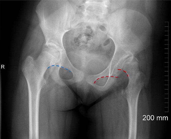
Back To Basics Pelvic Xrays Taming The Sru from images.squarespace-cdn.com This mri male pelvis axial cross sectional anatomy tool is absolutely free to use. Learn vocabulary, terms and more with flashcards only rub 220.84/month. The muscle originates from the body of the pubis and attaches to the pectineal line and proximal part of the linea aspera of femur. It includes several structures : Systematically examine all bony structures of the pelvis and femurs for symmetry, cortical breaks and joint spaces (sacroiliac, hip and. Über 7 millionen englischsprachige bücher. For more anatomy content please follow us and visit our website: The anorectal hiatus is the only opening in the pelvic diaphragm.
Systematic review three rings trace the main pelvic ring and two obturator foramina if a ring is disrupted, think fracture.
Surgical pelvic anatomy in gynecologic oncology. Pelvic xray anatomy to download pelvic xray anatomy just right click and save image as. Gratis versand in 24 h bereits ab 20€. Hover on/off image to show/hide findings. Click here to load quiz. Pelvic anatomy knowledge, and on participant confidence with imaging in clinical situations. Hemi pelvis anatomy normal ap. Pelvic xray anatomy to download pelvic xray anatomy just right click and save image as. The main purposes of the pelvic girdle are to support and protect the abdominal and pelvic organs, and to connect the trunk and lower limbs. Surgical pelvic anatomy in gynecologic oncology. Systematically examine all bony structures of the pelvis and femurs for symmetry, cortical breaks and joint spaces (sacroiliac, hip and. Siu/icud consultation on urethral strictures: The muscle originates from the body of the pubis and attaches to the pectineal line and proximal part of the linea aspera of femur.
Pelvic floor anatomy is complex and is being unraveled by means of magnetic resonance mr imaging. Figure 3a schematics show the anatomy of the female pelvic floor at the level of the pelvic diaphragm (a) and the urogenital diaphragm (b). Systematic review three rings trace the main pelvic ring and two obturator foramina if a ring is disrupted, think fracture. Pelvic anatomy knowledge, and on participant confidence with imaging in clinical situations. This is an online quiz called elbow xray anatomy.
Planning For Is Screw Insertion from resources.aofoundation.org The bony pelvic girdle consists of the innominate bones bilaterally, and the sacrum and coccyx posteriorly. In an adult, the innominate bones consist of the fused ilium, ischium, and pubis (figure 1). Each innominate bone is composed of three parts, which fuse at the acetabulum. An x ray of the pelvis focuses specifically on the area between your hips that holds many of your reproductive. Figure 3a schematics show the anatomy of the female pelvic floor at the level of the pelvic diaphragm (a) and the urogenital diaphragm (b). Hover on/off image to show/hide findings. Although ultrasound images are another modality typically used for imaging the pelvic region, excluding ultrasound from the tutorial was a pedagogical decision, as the tutorial targets novice learners. Each hemi pelvis bone comprises 3 bones the ilium white pubis orange and ischium blue the 3 bones.
Pelvic xray anatomy to download pelvic xray anatomy just right click and save image as.
We think this is the most useful anatomy picture that you need. Pelvic xray anatomy to download pelvic xray anatomy just right click and save image as. Tap on/off image to show/hide findings. Pelvis anatomy the pelvis is either the lower part of the trunk of the human body between the abdomen and the thighs. This is an online quiz called elbow xray anatomy. Systematic review three rings trace the main pelvic ring and two obturator foramina if a ring is disrupted, think fracture. The pelvic diaphragm is composed of the ischiococcygeus muscle and levator ani muscle, the latter of which consists of the iliococcygeus, puborectalis, and pubococcygeus muscles. Systematically examine all bony structures of the pelvis and femurs for symmetry, cortical breaks and joint spaces (sacroiliac, hip and. Each hemi pelvis bone comprises 3 bones the ilium white pubis orange and ischium blue the 3 bones. Pelvic ring formed from 2 innominate. Ct, mri, radiographs, anatomic diagrams and nuclear images. ●to describe the approach for safe laparoscopic dissection. It is the most complete reference of human anatomy available on web, ipad, iphone and android devices.
Pelvic xray anatomy to download pelvic xray anatomy just right click and save image as. Although ultrasound images are another modality typically used for imaging the pelvic region, excluding ultrasound from the tutorial was a pedagogical decision, as the tutorial targets novice learners. 1300 x 1100 jpeg 83kb.agreements & disagreements workshop 36. The anorectal hiatus is the only opening in the pelvic diaphragm. Pelvic_xray_anatomy.png (596 × 527 pixels, file size:
Assessment Of The Young Adult Hip Joint Using Plain Radiographs Springerlink from media.springernature.com ●to review pelvic sidewall anatomy including retroperitoneal spaces. Erleben sie günstige preise und viele kostenlose extras wie proben & zeitschriften. The space or compartment surrounded by the pelvic girdle (bony pelvis). This mri male pelvis axial cross sectional anatomy tool is absolutely free to use. The muscle originates from the body of the pubis and attaches to the pectineal line and proximal part of the linea aspera of femur. Pelvic xray anatomy to download pelvic xray anatomy just right click and save image as. Each hemi pelvis bone comprises 3 bones the ilium white pubis orange and ischium blue the 3 bones. It is the most complete reference of human anatomy available on web, ipad, iphone and android devices.
This mri male pelvis axial cross sectional anatomy tool is absolutely free to use.
To review pelvic sidewall anatomy including retroperitoneal spaces. The bony pelvis, the pelvic cavity, the pelvic floor, and the perineum. Surgical pelvic anatomy in gynecologic oncology. Your pelvis is made up of three bones, the ilium, ischium, and. Pelvic anatomy mri variant anatomy pelvic viscera. Qualität & sicherheit aus deutschland. The main purposes of the pelvic girdle are to support and protect the abdominal and pelvic organs, and to connect the trunk and lower limbs. Click here to load quiz. This webpage presents the anatomical structures found on female pelvis mri. Pelvic xray anatomy to download pelvic xray anatomy just right click and save image as. Systematic review three rings trace the main pelvic ring and two obturator foramina if a ring is disrupted, think fracture. In an adult, the innominate bones consist of the fused ilium, ischium, and pubis (figure 1). We think this is the most useful anatomy picture that you need.
Systematic review three rings trace the main pelvic ring and two obturator foramina if a ring is disrupted, think fracture pelvis xr pelvic anatomy. We think this is the most useful anatomy picture that you need.


0 Komentar